π‘¨π’„π’„π’†π’”π’”π’π’“π’š π‘΅π’‚π’—π’Šπ’„π’–π’π’‚π’“ π‘Ίπ’šπ’π’…π’“π’π’Žπ’†

𝑻𝒉𝒆 π‘·π’π’”π’•π’†π’“π’Šπ’π’“ π‘»π’Šπ’ƒπ’Šπ’‚π’ 𝑻𝒆𝒏𝒅𝒐𝒏 π‘°π’Žπ’‘π’π’”π’•π’π’“

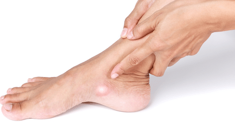
Medial arch pain and flatfoot deformities in runners and dancers are routinely diagnosed as Posterior Tibial Tendon Dysfunction (PTTD). But when eccentric calf raises aggravate the pain and a hard lump is visible on the inside of the foot, the patient likely has an extra bone hijacking the tendon.
​
Recent sports podiatry literature emphasizes that Accessory Navicular Syndrome (Os Naviculare) is a highly prevalent structural anomaly that chronically mimics tendinopathy and actively prevents standard arch rehabilitation from working.

What Is Accessory Navicular Syndrome?

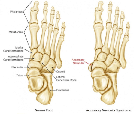
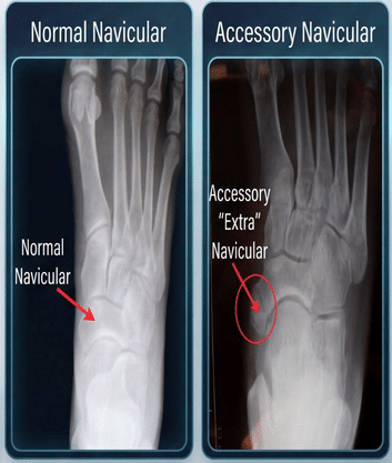
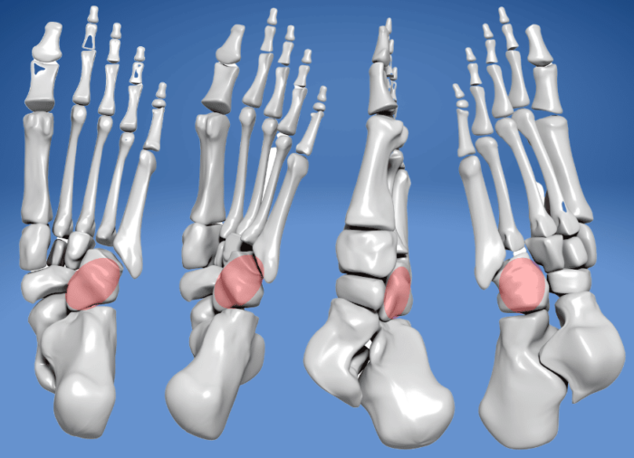
An accessory navicular is an extra piece of cartilage or bone located on the inner side of the foot, just above the arch. It is a congenital anomaly present in up to 14% of the population.
Β 
​Syndrome occurs when this extra bone becomes painful, typically because the posterior tibial tendonβ€”which is supposed to hold up the foot archβ€”abnormally attaches directly to this extra, unstable bone instead of its proper insertion point.

Pathophysiology

​When the posterior tibial tendon attaches to the accessory navicular, its mechanical line of pull is completely altered, losing its leverage to hold up the arch (leading to a flat foot).
Β 
​Furthermore, the fibrous joint (synchondrosis) connecting the extra bone to the main navicular bone is structurally weak. During high-impact activities (running, jumping) or direct trauma (like a tight ski boot rubbing the bump), the tendon violently pulls on the extra bone, literally trying to rip it away from the foot. This causes severe, chronic shear stress and inflammation at the fibrous interface.

Typical Pain Distribution

​Patients typically present with:
​‒ Deep, aching pain specifically on the medial (inner) aspect of the midfoot.
β€’ A highly visible, red, and swollen bony prominence protruding from the inside of the arch.
β€’ Pain that is drastically aggravated by wearing tight shoes (which rub the bump) or prolonged weight-bearing.

Key Clinical Signs

β€‹βœ”οΈ Focal Bony Tenderness: In true PTTD, the pain tracks along the tendon path behind the medial malleolus. In Accessory Navicular Syndrome, the pain and tenderness are located entirely and exactly on the bony bump itself.
βœ”οΈ Resisted Inversion Pain: Having the patient forcibly turn their foot inward against resistance will recreate the sharp pain at the bump, as the tendon pulls on the unstable bone.
βœ”οΈ MRI Bone Edema: 2025 imaging protocols show that symptomatic cases will exhibit intense bone marrow edema specifically at the synchondrosis (the junction between the extra bone and the main bone), proving it is a friction injury.

Why It Is Frequently Misdiagnosed

​Because it causes flat feet and medial arch pain, it perfectly mimics:
​‒ Posterior Tibial Tendinopathy (PTTD)
β€’ Spring Ligament Tears
β€’ Medial Column Foot Osteoarthritis
β€’ Tarsal Tunnel Syndrome

Evidence-Based Treatment Approaches

​You cannot strengthen a tendon that is attached to a structurally unstable, extra bone.
​Conservative management
β€’ Custom rigid orthotics with a deep heel seat and a medial “sweet spot” (a cutout specifically designed to offload pressure from the bony prominence).
β€’ Immobilization in a walking boot for 4 to 6 weeks to calm down acute flare-ups and allow the fibrous junction to scar down.
​Interventional options
β€’ If chronic pain persists, the surgical gold standard is the Kidner Procedure. This involves entirely excising (removing) the extra bone and surgically re-anchoring the posterior tibial tendon to its correct anatomical position on the main navicular bone, permanently restoring the mechanical leverage of the arch.

Clinical Takeaway

​If your “Posterior Tibial Tendinitis” patient has a visible bony bump on the inside of their foot that hurts to the touch, stop doing heavy calf raises. Palpate the prominence. Recognizing an Accessory Navicular changes the treatment plan from tendon loading to structural offloading and potential surgical removal.

You might also enjoy